Flow cytometry kit
|
product code
|
name registered by Yamasa
|
test count
|
|
|
telomere PNA Kit
|
K5327
|
TELOMERE PNA KIT/FITC
|
40
|
|
QIFIKIT
|
K0078
|
QIFIKIT
|
K5327 telomere PNA Kit
| This is a unique kit to detect telomere in vertebrate hematopoietic cells by fluorescence in situ hybridization (Flow-FISH) using flow cytometry. | |||||||||||||
Kit contents
|
|||||||||||||
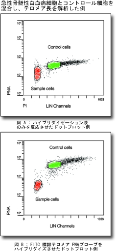 |
– A PNA probe with higher sensitivity and specificity than DNA probe is applied to flow cytometry.
– The PNA probe used in the kit does not recognize a subtelomeric sequence. Thus, unlike the conventional method of measuring a telomere restriction fragment (TRF), a telomere that contains no subtelomere can be detected. – After hybridization, post-hybridization procedures are minimally required, and washing with formamide is not needed. – Co-staining with PI allows telomere detection only with cells in the G0/1 phases. – By simultaneously measuring suitable control cells (e.g., 1301 cell), a relative telomere length can be determined. |
||||||||||||
K0078 QIFIKIT
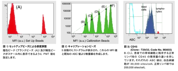
This is a unique kit with the concept of “quantification” added to the conventional flow cytometry analysis. A calibration curve is created using calibration beads coupled to five different amounts of antibody, allowing the quantification of antigen amounts as antibody-binding capacities (ABC: sites/cell) on cell surfaces of interest.
This kit can be used for measurements with all subclass IgG types of mouse monoclonal antibody.
In a study using flow cytometry, this kit allows the following examinations:
– Evaluation of the raised mouse monoclonal antibodies
– Changes in the amounts of antigen during the differentiation stages of blood cells
– Changes in the amounts of antigen after cell stimulation
– Search of relationship between pathological conditions and the amounts of antigen in leukemia and AIDS
Kit contents
| Vial1 | Setup beads | Blankbeads(A)+HighlevelBeads | 10 tests |
| Vial2 | Calibration beads | Beads(B,C,D,E,F) | 10 tests |
| Vial3 | FITC-labeled secondary antibody | Anti-mouse immunoglobulin goat antibody F (ab’) 2/FITC-labeled | 100 tests |
The kit includes a FITC-labeled secondary antibody. An optional RPE labeled secondary antibody (Code No. R0480) is available.