S100A9 levels in ulcerative colitis model rats
S100A9 is released from macrophages at the early stage of intestinal inflammation, and, therefore, is regarded as a useful marker of inflammatory bowel diseases. In addition, fecal S100A9 levels in ulcerative colitis model rats increase from the early stage, providing a useful marker for diagnosing inflammatory bowel diseases.
This page summarizes data on S100A9 levels in ulcerative colitis model rats. Neither reprint nor distribution is allowed without permission. If you have any questions, please contact the Marketing & MR Section of YAMASA Corporation. Some data are also listed on the product leaflet. Please download them as needed.
S100A9 expressions in the rectal tissues of DSS-induced ulcerative colitis model rats (UCR) were examined by fluorescence tissue staining. The model rats were prepared by administering dextran sulfate (DSS) to Wistar rats for seven consecutive days (with water administered from Day 8) (Figure 1). On Day 4 of administration, S100A8 and S100A9 were detected at comparable levels (yellow). However, on Day 6, only S100A9 increased (green). After the discontinuation of administration, S100A8 and S100A9 were detected at comparable levels.
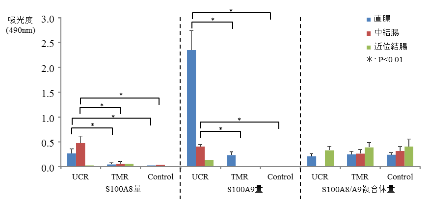
The complexes of S100A8/S100A9 and S100A8/S100A9 in rectal, middle, and proximal colonic tissues were measured on Day 6 of DSS administration (Figure 2). S100A9 was detected at a higher level in the rectal tissues of UCR than those of the control rats (control). S100A9 expression was suppressed in the rats (TMR) that simultaneously received an immunosuppressant, tacrolimus.
 Figure 1. Fluorescent tissue staining of the rectal tissues of ulcerative colitis model rats
Red: Anti-S100A8 antibody, Green: Anti-S100A9 antibody, Yellow: Merge
 Figure 2. S100A9 levels in the colonic tissue of ulcerative colitis model rats
UCR: DSS-induced ulcerative rat, TMR: DSS + tacrolimus-administered rat, Control: Water-administered control rats
(Data provided by Masao Ikemoto, a visiting professor from the Nagahama Institute of Bio-Science and Technology)
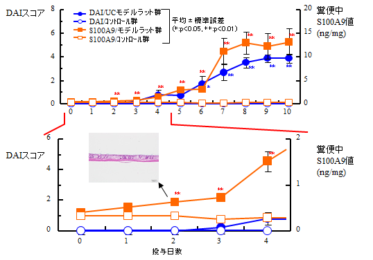
Feces were collected from the DSS-induced ulcerative colitis (UC) model and control rats to measure S100A9 levels using a kit. Simultaneously, their Disease Activity Index (DAI) scores, i.e., a total of diarrhea and blood stool scores (Figure 3), were compared.
On Day 2 after DSS administration, S100A9 levels were significantly higher in the UC group than in the control group (p<0.01). On Day 6, the DAI scores were significantly higher in the UC group than in the control group (p<0.05). Histological examination of the large intestine on Day 2 of DSS administration showed inflammatory symptoms, such as submucosal lymphocyte infiltration, suggesting that S100A9 reflects large intestinal inflammation from the early stage.
 Figure 3. Fecal S100A9 levels of ulcerative colitis model rats
(Source: The 42nd Annual Meeting of the Japanese Society of Clinical Toxicology (partially revised))
